

Computed tomography (CT)
What is computed tomography?
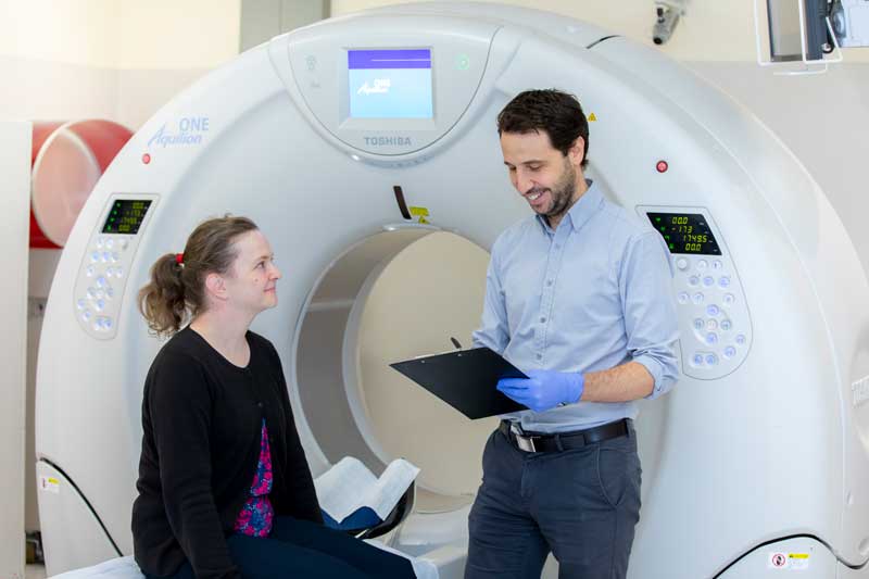
Computed tomography (CT) is a way of using X-rays to take pictures or images in very fine slices through the part of the body that the doctor has asked to be investigated. One way to think of it is of taking slices through a loaf of bread.
Modern scanners take more than one slice at a time. This may range from 4 to 320 slices and up to 640 slices for the most recent machines. This is referred to as ‘multi-slice‘ or ’multi-detector‘ technology.
Once the radiographer has taken the scan, these very thin slices can be put all together to reconstruct the loaf (or in this case your body). Once they are put back together, the radiographer can cut it into the slices in any direction that will help the radiologist (a doctor who has specialised in diagnostic imaging) to see the parts of the body that are of interest. Each scan is created specifically for the part of the body of interest and the condition that needs investigation. This may involve making several sets of pictures taken in different directions and also some 3-dimensional (3D) pictures.
With all of these different slices and 3D reconstructions, the radiologist will have a very detailed picture of the structures making up your body. This should help them to make a diagnosis (in other words, to understand the cause of your current problems) so that the right treatment can be planned as soon as possible.
